Medically reviewed by Dr Sin Yong · Last reviewed · 15 min read · Jump to questions
Medical-grade laser for idiopathic guttate hypomelanosis, vitiligo and post-inflammatory hypopigmentation — aiming to reactivate remaining melanocytes and improve skin tone uniformity where pigment can return. Singapore, Orchard Road.
White spots on the skin are usually hypopigmentation: idiopathic guttate hypomelanosis, post-inflammatory or procedure-related pigment loss, pityriasis versicolor or vitiligo. Treatment starts with dermoscopic assessment, because the key distinction is whether melanocytes are suppressed, which can recover, or lost, where repigmentation is limited; Dr Sin Yong's FSX Laser protocol is then matched to the type.
White spots on the skin — medically called hypopigmented lesions — occur when melanocytes (the cells that produce skin pigment) are damaged, absent or under-stimulated in a localised area. The appearance ranges from small scattered flat spots to larger irregular patches depending on the underlying cause.
In Singapore's tropical climate with high UV exposure, idiopathic guttate hypomelanosis (IGH) is particularly common — presenting as multiple small white spots on the forearms, shins and décolletage in adults from the age of 40 onwards. Post-inflammatory hypopigmentation following acne, eczema or previous procedures is also frequently seen.
Dr Sin Yong conducts a thorough dermoscopic assessment to accurately classify your white spots before selecting the appropriate treatment modality. Not all white spots respond to the same treatment — accurate diagnosis is the essential first step.

Small (2–5mm) flat white spots on sun-exposed areas — forearms, shins, shoulders. Linked to cumulative sun exposure and skin ageing; the exact cause is not known. Extremely common in Singapore adults over 40.
White patches following skin inflammation from acne, eczema, psoriasis or previous procedures. The melanocytes are suppressed rather than destroyed, making recovery more likely with treatment.
Fungal overgrowth (Malassezia) inhibits melanin production in affected skin. Typically presents as multiple small white patches on the trunk. Treated with antifungals; the pale patches can take weeks to months to even out after the yeast is cleared, and this does not mean treatment has failed.
Autoimmune destruction of melanocytes causing well-defined depigmented patches. Requires specialist management — laser can stimulate residual melanocyte activity at the lesion borders.
Depigmentation following aggressive chemical peels, cryotherapy or laser treatments by insufficiently calibrated devices. Response depends on whether melanocytes are suppressed or lost: suppressed pigment can recover, while delayed white spots after ablative resurfacing or frequent laser toning often respond slowly and incompletely.
Congenital hypopigmented patches present from birth. Melanocytes are present but dysfunctional. Response to laser is variable but can improve lesion appearance.
Dr Sin Yong uses the FSX (Flawless Smooth Skin) Laser protocol — a multi-wavelength laser approach — to address hypopigmentation by stimulating melanocyte proliferation and pigment migration. The aim is to encourage remaining melanocytes, at the lesion edge and in the hair follicle reservoir, to multiply and migrate into the pale skin; this is possible only where such melanocytes are still present.
Each lesion is assessed under dermoscopy to confirm the diagnosis and categorise lesion type. White spots caused by IGH, post-inflammatory hypopigmentation and vitiligo each receive a different laser parameter set — Dr Sin Yong does not use a generic "one size fits all" approach.
The FSX laser is applied to each white spot individually. The wavelength combination is selected based on lesion type and depth. The treatment aims to stimulate melanocytes in the follicular reservoir at the lesion border to proliferate and migrate centripetally — repopulating the hypopigmented area with functioning pigment-producing cells.
Sessions are repeated at intervals set at consultation. Where repigmentation occurs, it is gradual: small islands of pigment tend to appear first and may expand over later treatments. The number of treatments depends on lesion chronicity, size and location, and is reviewed at each visit.
Where melanocytes remain, pigment can return gradually as they repopulate the treated area. Change is judged over several months, not after a single visit.
Post-inflammatory hypopigmentation tends to respond well. IGH shows moderate improvement. Vitiligo response varies by location — facial lesions respond better than hands and feet.
For IGH and larger hypopigmented areas, FSX laser may be combined with topical agents (tacrolimus, retinoids) that support melanocyte activity. Dr Sin Yong designs a full protocol combining laser and home care for each patient.
Daily SPF 50 sunscreen is essential to maintain results and prevent new IGH lesions. Lifestyle sun protection — hats, UV clothing — is strongly recommended alongside treatment.
White spots or white patches on skin have several causes. The most common in Singapore are idiopathic guttate hypomelanosis (IGH) — small, flat white spots that appear on sun-exposed areas after years of UV exposure — pityriasis versicolor (a fungal skin condition), vitiligo, post-inflammatory hypopigmentation following acne or eczema, and chemical or laser-induced depigmentation. An accurate diagnosis is essential before treatment, as each condition responds to different therapies.
Yes — laser treatment for white spots depends on the underlying cause. For idiopathic guttate hypomelanosis and post-inflammatory hypopigmentation, the FSX (Flawless Smooth Skin) laser at Dr Sin Yong's practice uses a specific combination of wavelengths to encourage remaining melanocytes and improve pigment distribution where they are still present. For vitiligo, excimer-wavelength laser energy can reactivate melanocytes in depigmented patches. Results vary based on lesion type, skin tone and chronicity of the white spots.
Idiopathic guttate hypomelanosis and post-inflammatory hypopigmentation usually need a series of FSX laser treatments spaced a few weeks apart; the number depends on lesion type, size and how long the spots have been present. Vitiligo typically requires more sessions, and response varies by body location — facial lesions tend to respond better than acral (hands and feet) lesions. Dr Sin Yong will assess your lesions and give a realistic expectation of results during consultation.
The FSX laser protocol used by Dr Sin Yong is specifically calibrated for Asian and darker Fitzpatrick skin types (III–VI). The parameters are selected to keep the risk of post-inflammatory hyperpigmentation or paradoxical worsening low, though no laser removes that risk entirely. Patient safety and skin type appropriateness are assessed at every session.
Idiopathic guttate hypomelanosis (IGH) is a benign skin condition presenting as multiple small, well-defined white spots — typically 2–5mm — on sun-exposed areas such as the forearms, shins and shoulders. It is most common in adults over 40 and is associated with cumulative UV exposure. While medically harmless, IGH can be aesthetically distressing. Fractional laser, cryotherapy and topical retinoids or tacrolimus are the main options; studies are small, response varies, and IGH does not repigment on its own.
Usually because the laser has suppressed or reduced the pigment cells in that area. Lightening after inflammation often recovers with time and sun protection. Delayed white spots after ablative resurfacing or frequent laser toning can persist much longer, so the pattern needs assessing before anything else is done.
Some do and some do not. Where melanocytes are only suppressed, pigment often returns gradually over months with strict sun protection. Where melanocytes have been lost, as reported in guttate white spots after frequent laser toning, repigmentation is slow and often incomplete despite treatment.
No. IGH does not repigment by itself, and new spots can appear with age. Fractional laser, cryotherapy and topical retinoids or tacrolimus can improve the appearance of some spots, but studies are small and response varies. Daily sun protection helps slow the appearance of new spots.
The fee depends on the type and number of spots, the area treated, the laser settings chosen, whether topical agents are added and how many reviews are planned. A written quote is given at consultation after dermoscopic assessment, which also decides whether laser is advised at all for your type of white spot.
It can be worth considering where melanocytes are suppressed rather than lost, such as post-inflammatory hypopigmentation, and for selected idiopathic guttate hypomelanosis. It suits patients who accept partial, gradual change. It is the wrong tool for fungal patches, which need antifungals, and for areas where pigment cells have been lost.
No duration can be promised. Longevity depends on the type of white spot, how long it has been present, whether melanocytes remain, and sun exposure, because ultraviolet light can bring new spots. Maintenance with sun protection and topical care is part of the plan, and review timing is set at consultation.
Response is partial and variable, and some spots may not respond at all. Treatment is gradual and judged over months. Temporary redness is common, and darkening or further pigment loss can occur, especially in deeper skin tones. Several reviews are usually needed, and sun protection must continue to protect any change.
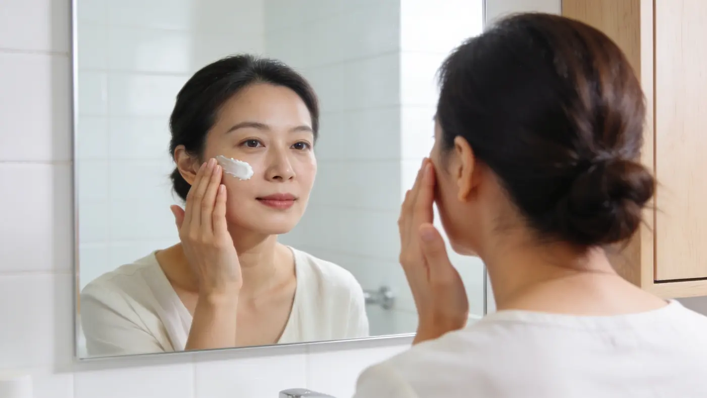
Dr Sin Yong assesses each hypopigmented lesion individually before recommending a treatment protocol. Available on Orchard Road, Singapore.
“Hypopigmentation is absent pigment, not scar. That distinction decides whether the skin can repigment at all — nothing applied to the surface rebuilds a melanocyte that has gone.”
Dr Sin YongOn why white marks are not one problem
Grimes PE, Bhawan J, Kim J, et al. Laser resurfacing-induced hypopigmentation: histologic alterations and repigmentation with topical photochemotherapy. Dermatologic Surgery 2001. source
Hang J, et al. Hypopigmentation following picosecond laser treatment for melasma: a case series. Lasers in Surgery and Medicine 2025. source
Hypopigmentation induced by frequent low-fluence, large-spot-size QS Nd:YAG laser treatments. source
Medically reviewed by Dr Sin Yong, MBBS (NUS), MRCS (Edinburgh) · Last updated 5 October 2026 · Editorial policy
| Option | What it does | What it cannot do | What to expect afterwards | Who it tends to suit |
|---|---|---|---|---|
| Post-inflammatory or laser-induced hypopigmentation | Melanocytes are suppressed after inflammation or a procedure; pigment can recover where they remain. | Delayed spots after ablative resurfacing or frequent laser toning often respond slowly and incompletely. | Change is judged over months with strict sun protection; an FSX Laser protocol may be matched. | Pale marks following acne, eczema or a procedure, after dermoscopic assessment. |
| Idiopathic guttate hypomelanosis (IGH) | Small flat white spots on sun-exposed skin, linked to cumulative sun exposure and ageing. | Does not repigment on its own, and new spots can appear with age. | Fractional laser, cryotherapy, retinoids or tacrolimus are options; studies are small and response varies. | Adults with scattered spots on forearms, shins or shoulders. |
| Vitiligo | Autoimmune loss of melanocytes, causing well-defined depigmented patches. | Needs specialist management; laser is not a stand-alone answer. | Laser can stimulate residual melanocyte activity at lesion borders; response varies by body site. | Well-defined depigmented patches needing specialist input. |
| Pityriasis versicolor | Fungal overgrowth (Malassezia) inhibits melanin production, typically on the trunk. | Laser does not treat the cause; it is treated with antifungals. | Pale patches take time to even out after the yeast clears; that is not treatment failure. | Multiple small pale patches on the trunk. |
| Dermoscopic assessment first | Classifies each white spot before any treatment is chosen. | Is not itself a treatment; no device is chosen before it. | Plan is matched to the type, with expectations set at consultation. | Anyone with white spots of unknown cause. |
People with long-standing patches where melanocytes have been lost, or with white spots caused by a fungal condition, tend to respond poorly, and laser is the wrong tool for them.
Expected effects include temporary redness, warmth and mild swelling in treated spots. Less commonly, laser can leave post-inflammatory darkening, particularly in deeper skin tones, or paradoxically worsen pigment loss, and no laser is free of that risk. Where melanocytes have been lost, little or no repigmentation may occur, and small studies of fractional laser and excimer light report partial, variable improvement. Blistering, infection and scarring are uncommon and relate to settings, skin condition and aftercare. Parameters are chosen for your skin tone and reviewed at each visit.
The fee depends on the type and number of white spots, the size and location of the area treated, the wavelength combination chosen, whether laser is combined with topical agents, and how many review visits the plan needs. A written quote is given at consultation after dermoscopic assessment. The consultation also decides whether laser is advised at all, since fungal causes need antifungal treatment first and some patches are unlikely to respond.
How quotes work at this practice: how we quote.
Many are idiopathic guttate hypomelanosis, post-inflammatory change or a fungal condition, and dermoscopic assessment separates them.
Laser can only encourage melanocytes that remain, so response depends on whether pigment cells are suppressed or lost.