Conditions · Face · Bumps & Growths

Seborrhoeic Keratosis (“Age Warts”)

Medically reviewed by Dr Sin Yong · Last reviewed · 10 min read · Assessed personally by Dr Sin Yong · Jump to questions

A seborrhoeic keratosis is a benign overgrowth of the skin's outermost cells — a waxy, warty plaque that looks pasted onto the surface, as if it could be picked off with a fingernail. It is not a wart, not contagious, not cancer, and not caused by anything you did. It is, overwhelmingly, just accumulated birthdays.

WhatsApp Dr Sin Yong →
Invited by Device Makers to Share His Expertise
International KOL for 14+ device brandsShared his clinical expertise with 500+ doctors across AsiaMBBS (NUS) · MRCS (Edinburgh) · MSc with Distinction (Queen Mary, London) · MSc (Cardiff)Every assessment personally by Dr Sin Yong

Seborrhoeic keratosis, often called an age wart, is a benign growth of the skin's surface cells: a waxy, sharply bordered, tan to dark brown plaque that looks stuck on. It is not a viral wart, not contagious and not cancerous, but melanoma can mimic it, so dermatoscopic diagnosis comes before removal by cryotherapy, curettage or laser.

Also called: age warts, seborrheic keratosis, 老人斑, 脂溢性角化

Key takeaways
  • Seborrhoeic keratosis, often called an age wart, is a benign growth of surface skin cells: a waxy, stuck-on, sharply bordered plaque.
  • It is not a viral wart, is not contagious and does not become cancer.
  • Melanoma and other pigmented skin cancers can mimic it, so dermatoscopic diagnosis comes before any removal.
  • Once confirmed benign, removal is elective, using cryotherapy, curettage, electrocautery or laser chosen by thickness and site.
  • New lesions can appear over the years, because the tendency is largely genetic and age-related.

Key Facts

What it is
Benign proliferation of keratinocytes — the skin's surface cells
The signature look
Waxy, 'stuck-on' plaque, tan to dark brown, sharply bordered, often slightly greasy-feeling
Who gets them
Most adults develop at least one by middle age; numbers increase with each decade
Where
Face, scalp, chest, back — anywhere except palms and soles
Malignant potential
None — but melanoma and pigmented skin cancers can mimic one, so diagnosis precedes removal
Removal modalities
Cryotherapy, curettage, electrocautery, ablative laser — quick, in-clinic
Who assesses this
A physician — any pigmented growth is diagnosed before destruction
Typical first step
Dermatoscopic confirmation it is benign; removal is then elective and quick

What does seborrhoeic keratosis look like?
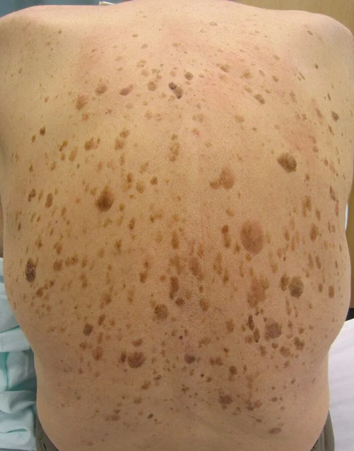
Multiple seborrhoeic keratoses — the 'stuck-on' age warts — across the back
Multiple seborrhoeic keratoses — the 'stuck-on' age warts — across the back. Image: James Heilman, MD, via Wikimedia Commons (CC BY-SA 3.0).
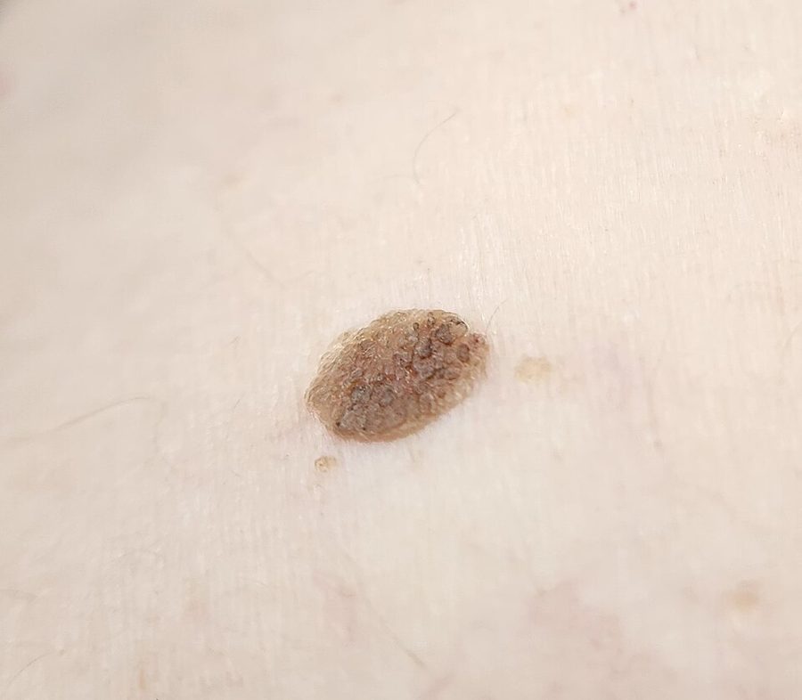
Close-up of a waxy, stuck-on keratosis
Close-up of a waxy, stuck-on keratosis. Image: Assafn, via Wikimedia Commons (CC BY-SA 4.0).

What are age warts, and why do they multiply?

Seborrhoeic keratoses are among the most common benign skin tumours in humans — reviews describe them as near-universal with ageing, driven by clonal changes in keratinocytes rather than by sun alone, though sun-exposed sites carry more [1]. They begin as flat tan patches and thicken over years into the classic waxy plaque. They run in families. They are not viral warts, despite the nickname — nothing spreads them, and picking one off (a popular home experiment) just brings it back from the base with a scab for company.

The one question that matters: is it definitely benign?

A seborrhoeic keratosis itself never becomes cancer. The clinical issue is the mimic: a melanoma can masquerade as a dark, irregular “SK”, and studies of excised lesions show a small but real rate of misdiagnosed malignancy hiding in presumed keratoses [1,2]. The rule Dr Sin Yong applies is absolute: any pigmented growth gets examined — dermatoscope where indicated — before anyone freezes, burns or lasers it. Destroying a lesion without diagnosis destroys the evidence. Features that fast-track review: rapid change, bleeding, an outlier that looks different from your other spots, or new pigmented lesions appearing in numbers.

What actually works for seborrhoeic keratosis?

Once confirmed benign, removal is elective and straightforward: cryotherapy for thin lesions, curettage or electrocautery for thicker ones, and ablative laser where precision or cosmetically sensitive sites call for it — the comparative literature supports all of these, chosen by lesion thickness and site [1,2]. On the face, technique matters more than modality: depth control determines whether the result is clear skin or a pale mark. Darker skin types need particular care with cryotherapy, which can leave lasting pigment change — one reason laser-based removal is often preferred in Singapore skin. Related surface concerns — flat brown patches and texture — are different problems with different tools.

Age warts are not the contagious kind

The lay name causes real confusion. A seborrhoeic keratosis is a benign overgrowth of keratinocytes with no viral component: it cannot be caught, cannot be passed to anyone else, and does not seed to other parts of the body. A viral wart, caused by human papillomavirus, does all three.

This matters before anything is treated, because the two need different handling and a lesion assumed to be an age wart should first be confirmed as one. The comparison is set out in full in warts versus age warts.

What doesn't work for seborrhoeic keratosis?

Creams — no topical dissolves a thick keratinocyte plaque reliably; the “SK removal” products sold online mostly irritate. Picking — it regrows from the base. Home freezing kits — uncontrolled depth on an undiagnosed pigmented lesion is the worst combination in dermatology. And ignoring the changing one because “it's just an age wart” — the mimic problem is exactly why the changing one earns a doctor's eyes.

If the lesion in question is a true mole rather than a seborrhoeic keratosis, Dr Sin Yong’s dermatoscopy-first mole removal protocol applies — the mole is scoped and assessed before any removal method is chosen.

“A seborrhoeic keratosis never turns into cancer — but cancer occasionally dresses as one, which is why diagnosis comes before destruction.”

— Dr Sin Yong

Dr Sin Yong’s Viewpoint

My rule for seborrhoeic keratoses is absolute: diagnosis before destruction. The lesion itself never turns malignant — but melanoma occasionally dresses as one, and the changing, outlier 'age wart' is the one that earns my dermatoscope. Once confirmed benign, removal is quick, elective and satisfying.

Prefer Not to Use WhatsApp?

Use the short enquiry form instead →

Leave your name, mobile number and email and the clinic will contact you directly. No obligation.

Your details are delivered directly to Dr Sin Yong's clinic team.

Thank you — your enquiry has been received. The clinic will contact you during opening hours (Mon–Fri 10am–8pm, Sat 11am–3pm).

Prefer a preliminary view first? You can also WhatsApp a photo of the area for Dr Sin Yong to review before you decide on a visit.

Which type do you have?

Seborrhoeic keratosis and its look-alikes
Type / look-alikeHow to recognise itWhat it needsWhat does not work
Seborrhoeic keratosisWaxy, stuck-on plaque, tan to dark brown, sharply borderedDermatoscopic confirmation, then elective removal if wantedCreams, picking and home freezing kits
Viral wartRough lesion that can spread to other sites and other peopleConfirmed diagnosis and wart-specific treatmentHandling it as an age wart
Melanoma mimicDark, irregular or changing lesion unlike your other spotsDermatoscopic examination and, where needed, biopsy firstFreezing, burning or lasering without a diagnosis
Flat brown patch (lentigo)Flat, evenly pigmented patch without a thick waxy surfaceDiagnosis and a pigment-directed planTreating it as a thick keratosis
Mole (naevus)Pigmented spot that is flat or raised, assessed by dermatoscopeDermatoscopy before any removal method is chosenHome removal or destruction without diagnosis

What does not work, and why

When to see a doctor

Myths we hear in clinic

“Age warts are contagious, like viral warts.”

A seborrhoeic keratosis has no viral component and cannot be passed to anyone or spread to other parts of your body.

“If it looks like an age wart, it is one.”

The stuck-on look is reassuring but not diagnostic, because some melanomas imitate it, so examination settles it.

Questions Patients Actually Ask

Are seborrhoeic keratoses contagious?+

No. Despite the 'wart' nickname they are not viral — nothing spreads them between people or across your own skin.

Why am I suddenly getting so many?+

Numbers rise with age and family tendency. A genuinely sudden eruption of many new lesions is uncommon and worth a medical review rather than assumption.

Can I tell an SK from a melanoma myself?+

Not reliably — some melanomas imitate the waxy look convincingly. The stuck-on appearance is reassuring but not diagnostic; examination takes minutes and settles it.

Does removal hurt?+

Removal is done under local anaesthetic or with brief, tolerable discomfort depending on modality and size. Most patients find it far easier than expected.

Will they come back after removal?+

A properly removed lesion rarely regrows, but new ones can appear elsewhere over the years — the tendency is constitutional.

Is there any way to prevent them?+

No proven prevention exists; they are largely genetic and age-driven. Sun protection is sensible for many reasons but only partially influences these.

What causes seborrhoeic keratosis?+
Seborrhoeic keratosis arises from clonal changes in the skin's surface keratinocytes. It is largely genetic and age-related, so many adults develop at least one and numbers rise with each decade. Sun exposure plays a part on exposed sites but is not the whole story. It is not caused by a virus, and it cannot be caught or spread.
Can seborrhoeic keratosis be fully removed?+
Once a lesion is confirmed benign, it can be removed in clinic by cryotherapy, curettage, electrocautery or laser, chosen by thickness and site. A properly removed lesion rarely regrows, but new ones can appear elsewhere because the tendency is constitutional. Depth control on the face affects how the skin looks afterwards, and no outcome is promised.
Which treatment suits seborrhoeic keratosis?+
Diagnosis comes first, because melanoma can mimic a seborrhoeic keratosis. After dermatoscopic confirmation, removal is optional. Thin lesions suit cryotherapy, thicker ones curettage or electrocautery, and laser suits sites where precision matters. Skin type affects the choice, since cryotherapy can leave pigment change in darker skin.
How much does seborrhoeic keratosis treatment cost in Singapore?+
The cost follows the plan. It depends on the number, thickness and size of the lesions, their site, the method chosen, the device and consumables used and whether several lesions are treated in one visit. A written quote is given at consultation after dermatoscopic assessment, and the consultation decides whether removal is advised at all.

References

  1. Recent Advances in Managing and Understanding Seborrheic Keratosis — F1000Research (PMC).
  2. Efficacy and Safety of Topical Treatments for Seborrheic Keratoses: A Systematic Review — Journal of Dermatological Treatment (PubMed).
  3. Seborrhoeic Keratosis — DermNet NZ.

Medically reviewed by Dr Sin Yong, MBBS (NUS), MRCS (Edinburgh) · Last updated 7 September 2026 · Editorial policy

Follow Dr Sin Yong
InstagramTikTokYouTubeThreadsXLinkedInFacebook
Dr Sin Yong · Every assessment personally performed
WhatsApp Enquiry