Medically reviewed by Dr Sin Yong · Last reviewed · 22 min read · Doctor-performed, never delegated · Jump to questions
Dr Sin Yong · MBBS (NUS) · MRCS (Edin) · MSc Aesthetic Medicine with Distinction (Queen Mary, London) · MSc Practical Dermatology (Cardiff) · International KOL
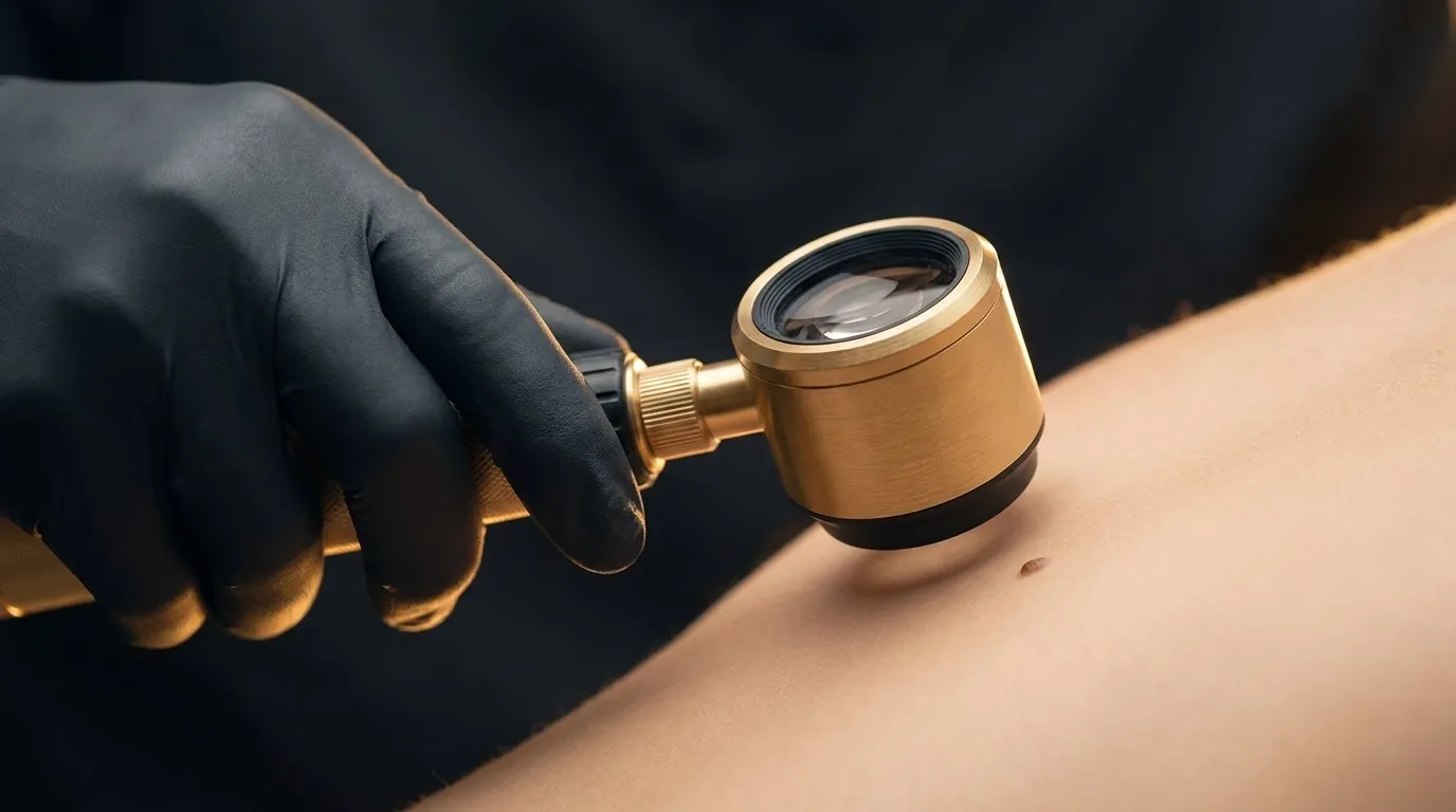
Every mole at Dr Sin Yong's practice is examined under a dermatoscope before anything touches it. Dermatoscopy reveals the pigment pattern, borders and vascular structures the naked eye cannot see — so a mole is only removed once it has been properly assessed, and removed with the method that suits its type, depth and location.
Mole removal in Singapore should begin with a dermatoscope examination. At Dr Sin Yong's practice each mole is scoped first; benign moles are removed by CO2 laser, radiofrequency or shave under local anaesthesia, while any mole with atypical features is excised for histology or referred. The method follows the mole's type, depth and location.
Also called: naevus removal, 去痣, 除痣, ほくろ除去
A dermatoscope is a polarised magnification instrument that lets a trained physician see beneath the surface glare of the skin — the pigment network, globules, dots, streaks and blood-vessel patterns that distinguish an ordinary mole from one that deserves closer attention. Published meta-analysis shows dermatoscopy meaningfully improves the accuracy of melanocytic-lesion assessment compared with naked-eye examination alone.
Once a mole has been scoped and assessed as suitable for cosmetic removal, its type decides the technique. Flat mole removal and raised mole removal are different procedures: a flat junctional mole responds to precise CO2 laser mole removal in fine layers, while a raised dermal mole may suit radiofrequency or shave removal instead. A raised dermal mole behaves differently from a flat junctional one; a mole on the nasal fold needs a different approach from one on the back. Facial mole removal demands particular finesse — a mole on the nose, eyelid or lip margin heals in full view, so Singapore mole removal patients are right to ask how the site will look afterwards, not just how the mole comes off. Matching lesion to method is what determines how cleanly the site heals — the heart of careful aesthetic mole removal. Where dermatoscopy raises any concern, the priority changes — the mole is not ablated; it is excised in a manner that preserves tissue for histological examination, or referred appropriately.
A mole is a diagnosis, not just a blemish. Burning or cutting a mole off without dermatoscopic assessment destroys the very information a doctor needs — and on the rare occasion a lesion is not what it seems, that information matters. Scope first. Then remove.

A dermatoscope examines the mole at roughly tenfold magnification under cross-polarised light, removing surface glare to reveal sub-surface pigment structure.
Relative diagnostic odds ratio for melanoma assessment with dermatoscopy versus naked-eye examination in the Vestergaard 2008 meta-analysis of clinical studies.
Wavelength of the CO2 laser used for layered ablation of scoped, benign moles — absorbed by water in tissue for micron-level control.
Typical operating frequency of radiofrequency removal, which shaves and coagulates in the same pass.
The classic ABCDE diameter threshold above which a mole warrants professional dermatoscopic assessment rather than casual removal.
Depth range separating a flat junctional mole (near the epidermis, ~0.1 mm) from a raised dermal mole extending millimetres deeper — the anatomy that decides the removal method.
Each mole is examined under the dermatoscope. Structure, symmetry, border, pigment pattern and vessels are assessed — not just its surface appearance.
Benign-pattern moles are mapped for cosmetic removal. Any lesion with atypical features is redirected to a biopsy-preserving pathway instead of ablation.
Laser ablation, radiofrequency or shave removal is chosen according to the mole's type, depth and location — the decision the scope makes possible.
The mole is removed precisely, layer by layer where relevant, respecting the surrounding skin.
Where assessment calls for it, removed tissue is sent for histological examination rather than vaporised.
Wound-care guidance protects the healing site, and review confirms it has settled as expected.
The ABCDE rule is the internationally used screen for moles that deserve professional assessment rather than casual removal:
One half does not match the other
Irregular, blurred or notched edges
More than one shade within a single mole
Larger than about 6 mm, or growing
Changing in size, shape, colour or sensation
Any of these features is a reason to scope — never a reason to reach for an over-the-counter mole pen or a beauty-salon cautery. The dermatoscope settles what the surface cannot.
“Truly ‘scarless’ mole removal is a marketing phrase, not a medical one.”
— Dr Sin Yong
Laser mole removal, radiofrequency, shave removal or mole removal surgery — no single method is right for every mole. The dermatoscopic assessment decides which column applies to yours.
| CO2 laser ablation | Radiofrequency | Shave removal | Surgical excision | Monitor only | |
|---|---|---|---|---|---|
| What it is | Focused CO2 laser vaporises the mole in fine layers | High-frequency energy shaves and seals in one motion | The raised portion is shaved level under local anaesthesia | The full lesion is removed and sent for histology | The mole stays; it is photographed and re-scoped over time |
| Suited for | Small facial and body moles with benign dermatoscopic pattern | Raised dermal moles; delicate locations | Protruding moles where flatness is the goal | Any mole with atypical features on dermatoscopy | Benign moles the patient prefers to keep |
| Anaesthesia | Topical or local | Local | Local | Local | None |
| Typical downtime | A small scab for about 1–2 weeks | A small scab for about 1–2 weeks | A flat mark that fades over weeks | Sutured wound; sutures out in about 1–2 weeks | None |
| Histology available | Limited — tissue is vaporised | Limited | Yes — shaved tissue can be sent | Yes — the entire lesion is examined | Not applicable |
| The role of the scope | Confirms benign pattern before ablation | Confirms benign pattern before ablation | Confirms the mole suits a shave | The scope is what triggers this pathway | The scope is what justifies leaving it alone |
Indicative comparisons for patient education — the appropriate method, healing course and any need for histology are determined at consultation for each individual mole.
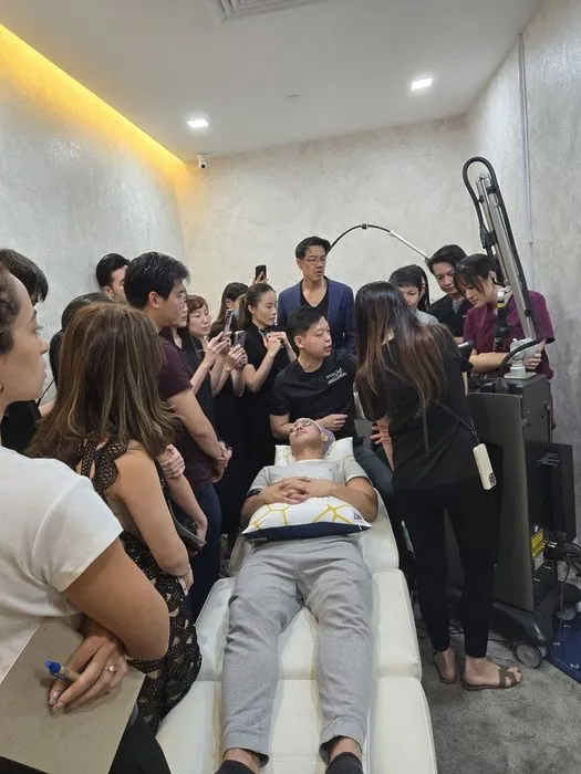
Precision energy work is exactly that — precision. The physician assessing your mole is the one international device manufacturers invite to share his clinical expertise with other doctors on laser and energy-based treatments:


“Do not remove your mole until you have scoped it.”
— Dr Sin Yong
Flat mole removal Singapore and raised mole removal are different procedures, and conflating them is how people end up disappointed. A raised mole sits above the skin plane and can often be taken down to that plane; a flat mole is pigment within the skin, so removing it means treating into the dermis rather than shaving anything off. The methods, the healing and the mark left behind all differ.
Which brings up the mole removal scar question, which deserves a direct answer: any removal leaves something. The realistic objective is trading a visible mole for a mark that is flatter, paler and less noticeable than what it replaced — not the absence of any evidence. Anyone promising otherwise is describing a different lesion.
The prior step is not cosmetic. Every mole is examined under a dermatoscope before any discussion of removal, because a lesion that should go to histology must not be treated as a cosmetic item.
A wart and a mole are different lesions, so the first step in wart removal is confirming that the growth is a wart at all. A mole is a cluster of pigment cells. A viral wart is a rough, thickened growth caused by the human papillomavirus, often with tiny dark dots, and it can spread. A seborrhoeic keratosis, often called an age wart, is a stuck-on, waxy growth that is neither viral nor a mole.
The distinction changes what happens next. Moles go through the dermatoscopy-first pathway described above. Seborrhoeic keratoses are removed by methods suited to their thickness and site, and have their own page. Viral warts follow a different logic: common approaches include salicylic acid preparations and cryotherapy, and persistent warts, or warts on the soles or genitals or in people with lowered immunity, are usually managed by a GP or dermatologist.
A growth that looks like a wart but is pigmented, bleeding, changing or unlike your other lesions should be examined with a dermatoscope before anything is frozen, burnt or cut, because some skin cancers can resemble a wart.
Get a mole checked when it is new in adulthood, changing, or different from your other moles. A mole that grows, darkens unevenly, changes shape, itches, bleeds or crusts without injury should be examined promptly rather than watched at home. The ABCDE features above are the formal screen; two further signs are worth knowing.
The first is the 'ugly duckling' sign: a mole that looks unlike the rest of your moles deserves attention even if it passes each ABCDE test. The second is location. In Asian skin, melanoma is proportionally more often found on the palms, soles and under the nails, so a new or changing dark spot on the sole, or a dark streak running along a nail, should be examined rather than dismissed as a bruise or a stain.
Examination does not mean removal. Most moles that are checked are benign and can be left alone, or removed for cosmetic reasons later. What changes is that the decision is made with information: the dermatoscopic pattern, photographs for comparison at review and, where anything is uncertain, excision with histology or referral to a dermatologist.
Most red moles are cherry angiomas: small, benign clusters of dilated blood vessels rather than moles at all. They are bright red to purple, flat or dome-shaped, become more numerous from early adulthood and are common on the trunk and arms. They contain no pigment cells and do not turn into melanoma.
Because they are vascular, they are not treated like a pigmented mole. Small angiomas are commonly removed with radiofrequency or electrocautery, or treated with a vascular laser, and the method depends on size, site and how raised the lesion is. A small mark can remain where a larger angioma has been treated, and new ones may continue to appear over the years.
Not every red spot is a cherry angioma. A red lump that appeared quickly, bleeds easily or keeps re-forming may be a pyogenic granuloma, and rarely a skin cancer can be pink or red with little visible pigment. That is why a red lesion is still examined under the dermatoscope before removal, and why anything atypical goes for histology rather than being cauterised away.
Cosmetic removal is not the right step for a mole with atypical features on dermatoscopy, a mole that is changing, bleeding without injury or unlike your others, or any lesion whose diagnosis is uncertain. Those are excised for histology or referred, rather than lasered, because ablation destroys the tissue a pathologist would need.
Other people need extra care or a different plan. A personal or family tendency to keloid or thickened scars matters, particularly on the chest, shoulders, upper back and jawline. Blood-thinning medicines, a bleeding tendency, poorly controlled diabetes or slow healing, and allergy to local anaesthetic are reviewed first. Active infection, eczema or sunburn at the site is allowed to settle. In pregnancy a purely cosmetic removal is usually deferred, while a mole that is changing is still examined promptly. Recently tanned skin is assessed for the risk of pigment change at the healed site. Whether, when and how a mole is removed is decided at consultation.
Removing a scoped, benign mole under local anaesthesia is a routine procedure, but every method carries risks. The most predictable is a mark: any removal that reaches the mole leaves a scar, usually flatter and paler than the mole, and less often slightly depressed, raised or darker. Post-inflammatory hyperpigmentation is more common in Asian skin and is reduced by sun protection while the site heals; a lighter patch can also occur.
Other risks include infection, bleeding, slow healing, and keloid or thickened scarring in people prone to it. Re-pigmentation is more likely when deeper mole cells are left behind: in one study of 204 common moles removed by shave excision, clinical or dermatoscopic signs of recurrence were seen at 19.6% of sites at three months.
The most serious risk is diagnostic rather than technical: treating a lesion that should have gone to histology. That is why every mole is scoped first, the method is matched to its depth and site, and any regrowth is re-examined rather than simply treated again.
The sequence below describes phases, not a timetable: how quickly each passes depends on the settings used, the area and the person, and is discussed at consultation.
People expecting a completely mark-free result, or wanting a changing or suspicious mole simply taken off without examination, tend to be poorly served by cosmetic removal.
The fee depends on how many moles are treated, their size, depth and site, and the method chosen, whether CO2 laser, radiofrequency, shave or excision. It also depends on whether tissue is sent for histology and whether removal is combined with another treatment. A written quote is given at consultation after dermatoscopic assessment, and the consultation decides whether removal is advised at all.
How quotes work at this practice: how we quote.
Burning or cutting a mole off without dermatoscopy destroys the information a doctor needs, so scope first and remove second.
Laser ablation suits moles with a clearly benign dermatoscopic pattern, while atypical lesions need excision with histology.
Because the surface tells only part of the story. Dermatoscopy shows the pigment network and vessel pattern beneath the surface, which is how a physician distinguishes an ordinary benign mole from a lesion that should be excised and examined instead of vaporised. It takes minutes, and it is the difference between removing a mole and removing information.
The plan changes. An atypical lesion is not lasered off — it is removed in a way that preserves the tissue for histological examination, or referred for specialist evaluation. This is precisely why scoping comes before removal, never after.
That is decided at assessment. Small benign moles are commonly treated with mole removal laser techniques (CO2) or radiofrequency; protruding moles may suit shave removal; lesions needing histology are excised. Type, depth and location of the mole drive the choice.
The area is numbed with topical or injected local anaesthesia before removal, and comfort is checked before the mole is treated. Mild tenderness at the site afterwards is common, and aftercare is explained on the day.
Any removal leaves a mark that matures over weeks to months; the aim of matching method to mole is to keep that mark as inconspicuous as the lesion and location allow. Wound care instructions — and sun protection over the healing site — meaningfully influence the final result.
No — and that is the point of scoping first. Laser ablation is appropriate for moles with a clearly benign dermatoscopic pattern. A lesion with atypical features needs excision with histology, not vaporisation.
A small scab typically forms and separates over one to two weeks, with residual redness fading over the following weeks. Healing time varies with the mole's size, depth, the method used and the site on the body.
A completely removed mole does not usually recur, though deeper dermal components can occasionally re-pigment and may need a touch-up assessment. Any regrowth should itself be re-scoped rather than assumed harmless.
No. No matter what a mole removal cream Singapore retailer promises, creams, corrosive pens and salon cautery all remove tissue without assessment, frequently scar, and destroy the possibility of histological examination. A mole should be assessed by a physician with a dermatoscope before anyone removes it — that is the standing rule of this practice.
Multiple scoped, benign moles can commonly be treated in a single sitting. The number is agreed at consultation based on their sites and the aftercare involved.
The mole removal price in Singapore depends on the removal method, and the number, size and location of the moles being treated — so the mole removal Singapore cost for your case is quoted individually after the dermatoscopic assessment at consultation. Fees are always confirmed with you before any procedure is done.
Truly 'scarless' mole removal is a marketing phrase, not a medical one — any removal that reaches the mole leaves some mark. What careful technique can do is keep that mark small, flat and inconspicuous by matching the method to the mole's type, depth and location, and by protecting the healing site afterwards.
Yes. A flat mole sits at or near the junction of the skin layers and is usually removed by CO2 laser in fine, controlled layers, while a raised dermal mole extends deeper and may respond better to radiofrequency or shave removal. This is one more decision the dermatoscope settles before treatment begins.
Question not answered here? Ask on WhatsApp, use the enquiry form, or browse 100 questions patients ask.
Mole removal in Singapore at Dr Sin Yong's practice always begins with dermatoscopic examination. Each mole is scoped and assessed before removal; benign moles are removed by CO2 laser, radiofrequency or shave technique under local anaesthesia, while any mole with atypical dermatoscopic features is excised with histological examination or referred appropriately. This information is educational and is not a substitute for a medical consultation.
Medically reviewed by Dr Sin Yong, MBBS (NUS), MRCS (Edinburgh) · Last updated 7 October 2026 · Editorial policy
Dermatoscope-first mole assessment, and removal matched to your mole — laser, radiofrequency, shave or excision.
Replies within clinic hours: Mon–Fri 10am–8pm, Sat 11am–3pm · How consultations work · Prefer not to use WhatsApp? · Treatment finder
WhatsApp Dr Sin YongLeave your name, mobile number and email and the clinic will contact you directly. No obligation.